Wako 血清素相关抗体 Serotonin-related antibody
Other psychiatric disorders
Serotonin-related antibody
- Anti-mouse 5-HT1A receptor, rat monoclonal antibody (4A6)
Anti-mouse 5-HT2C receptor, rat monoclonal antibody (6D2) - Anti-mouse serotonin transporter, rat monoclonal antibody (R5-3-2)
- Product List
- Related Information
Anti-mouse 5-HT1A receptor, rat monoclonal antibody (4A6)
Anti-mouse 5-HT2C receptor, rat monoclonal antibody (6D2)
5-HT1Areceptor and 5-HT2C receptor are G protein-coupled receptors activated by serotonin (5-HT). Both are localized mainly in the central nervous system, and their functions controlling memory, food intake, sleep, pleasure, and anxiety, etc. Anxiolytics and antipsychotics acting on these receptors have been developed, and they are attracting attention of researchers as novel targets for drug discovery.
This product is a rat monoclonal antibody established by DNA immunization and specifically recognizes native forms of 5-HT1A receptor and 5-HT2C receptor.
抗小鼠 5-HT 1A受体、大鼠单克隆抗体 (4A6)
抗小鼠 5-HT 2C受体、大鼠单克隆抗体 (6D2)
5-HT 1A受体和5-HT 2C受体是由5-羟色胺(5-HT) 激活的G 蛋白偶联受体。两者都主要位于中枢神经系统,具有控制记忆、食物摄取、睡眠、愉悦和焦虑等的功能。已经开发出作用于这些受体的抗焦虑药和抗精神病药,它们作为新靶点正在引起研究人员的关注。药物发现。
本品是通过DNA免疫建立的大鼠单克隆抗体,特异性识别天然形式的5-HT 1A受体和5-HT 2C受体。
Features
- Compatible with immunohistochemical staining
- Specifically recognizes native forms of 5-HT1Areceptor or 5-HT2C receptor
- Rat monoclonal antibodies established by DNA immunization
特征
- 与免疫组化染色兼容
- 特异性识别天然形式的 5-HT 1A受体或 5-HT 2C受体
- DNA免疫法建立的大鼠单克隆抗体
Description
| Anti 5-HT1A receptor antibody | Anti 5-HT2C receptor antibody | |
|---|---|---|
| Clone No. | 4A6 | 6D2 |
| Applications | Immunohistochemistry (1:100-2,000) Flow cytometry(1:100-1,000) |
Immunohistochemistry (1:200-10,000) Flow cytometry(1:100-1,000) |
| Subclass | Rat IgG2b・κ | Rat IgG2a・κ |
| Cross-reactivity | Mouse ※not tested for other animal species | |
| Antigen | Vector expressing mouse 5-HT1A receptor | Vector expressing mouse 5-HT2C receptor |
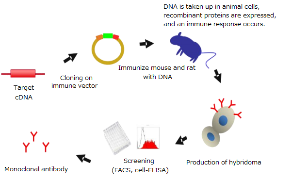
DNA immunization
What is DNA immunization?
DNA immunization is a technique for generating antibodies against target proteins by using antigen proteins expressed in the animal body with expression vectors containing the target genes.
DNA免疫
什么是 DNA 免疫?
DNA免疫是一种利用在动物体内表达的抗原蛋白和含有靶基因的表达载体产生针对靶蛋白的抗体的技术。
Features of DNA immunization
- Yields Antibodies specifically recognizing native forms of proteins
- Suitable for preparation of antibodies recognizing membrane proteins that were difficult to prepare by conventional methods
DNA免疫的特点
- 产生特异性识别天然蛋白质形式的抗体
- 适用于制备识别常规方法难以制备的膜蛋白的抗体
Principles of DNA immunization
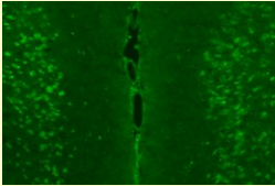
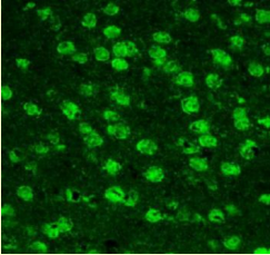
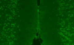
Usage example – Immunohistochemical staining
5-HT1Areceptor antibody
-

-
-
Localization in the cell body of prefrontal neurons was confirmed.
This localization is consistent with the site highly expressing mRNA for 5-HT1A.
5-HT2C receptor
-
-
-
Localization in the cell body of prefrontal neurons was confirmed.
This localization is consistent with the site highly expressing mRNA for 5-HT2C.
Experimental conditions
Specimen: Prefrontal area of the brain from 10-week-old wild-type mice
Sections: 12 µm-thick frozen sections
Activation condition: Microwave treatment in a citrate buffer (pH 7.0) for 10 minutes
Antibody concentration: 1 µg/mL
实验条件
标本:来自 10 周龄野生型小鼠的大脑前额叶区域
切片:12 µm 厚的冰冻切片
激活条件:在柠檬酸盐缓冲液 (pH 7.0) 中微波处理 10 分钟
抗体浓度:1 µg/mL
Usage example – Flow cytometry
5-HT1A receptor antibody
-
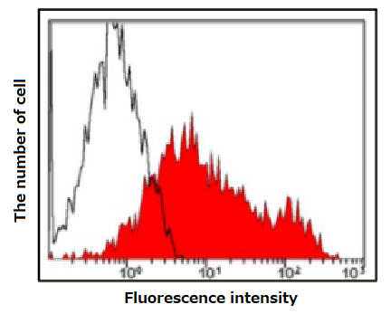
Open area: 293T cells
Red area: 293T cells expressing 5-HT1A receptorAntibody concentration: 1 µg/mL -
An obvious shift was observed only for cells expressing 5-HT1A.
Anti 5-HT2C receptor antibody
-
This product 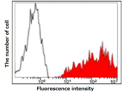
Open area: 293T cells
Red area: 293T cells expressing 5-HT2C receptorAntibody concentration: 1 µg/mL -
Competitor 
-
A clearer shift was observed for cells expressing 5-HT2C, compared with the competitor.
Usage example – Immunohistochemical staining of the site expressing mRNA for 5-HT1A
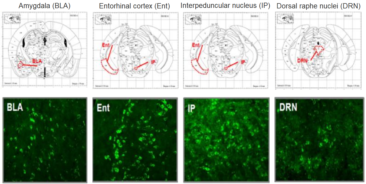
Positive signals were noted in amygdala (BLA), entorhinal cortex (Ent), interpeduncular nucleus (IP), and dorsal raphe nuclei (DRN).
This localization is consistent with the site highly expressing mRNA for 5-HT1A.
Experimental conditions
Specimens: Various areas of the brain from 10-week-old wild-type mice
Sections: 12 µm-thick frozen sections
Activation condition: Microwave treatment in a citrate buffer (pH 7.0) for 10 minutes
Antibody concentration: 1 µg/mL
Usage example – Immunohistochemical staining of the site expressing mRNA for 5-HT2C receptor
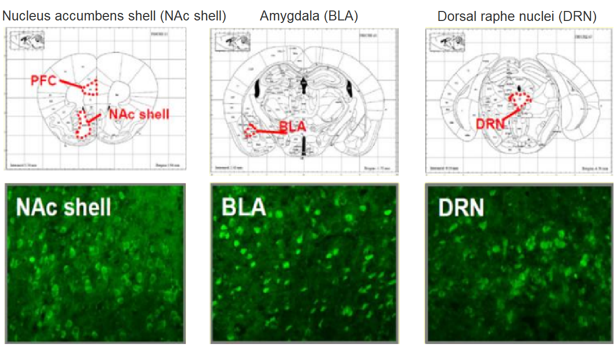
Positive signals were noted in nucleus accumbens shell (NAc shell), amygdala (BLA), and dorsal raphe nuclei (DRN).
This localization is consistent with the site highly expressing mRNA for 5-HT2C.
Experimental conditions
Specimens: Various areas of the brain from 10-week-old wild-type mice
Sections: 12 µm-thick frozen sections
Activation condition: Microwave treatment in a citrate buffer (pH 7.0) for 10 minutes
Antibody concentration: 1 µg/mL
All immunohistochemical staining image data presented above are courtesy of Dr. Matsuda, Graduate School of Pharmaceutical Sciences, Osaka University in Japan and Dr. Takuma and Hasebe of Graduate School of Dentistry, Osaka University in Japan.
实验条件
标本:10 周龄野生型小鼠大脑的各个区域
切片:12 µm 厚的冰冻切片
激活条件:在柠檬酸盐缓冲液 (pH 7.0) 中微波处理 10 分钟
抗体浓度:1 µg/mL
以上所有免疫组化染色图像数据均由日本大阪大学药学研究生院 Matsuda 博士和日本大阪大学牙科研究生院的 Takuma 和 Hasebe 博士提供。
Anti-mouse serotonin transporter, rat monoclonal antibody (R5-3-2)
Serotonin transporter is a 12-transmembrane protein. It incorporates extracellular serotonin in the brain into the presynapse to modulate serotonin levels. With its reported involvement in sleep, fear, and anxiety, serotonin transporter has been investigated as a target of antidepressants.
抗小鼠血清素转运蛋白、大鼠单克隆抗体 (R5-3-2)
血清素转运蛋白是一种 12 跨膜蛋白。它将大脑中的细胞外血清素结合到突触前以调节血清素水平。据报道,血清素转运蛋白与睡眠、恐惧和焦虑有关,已被研究作为抗抑郁药的目标。
特征
- 与免疫组化染色兼容
- 通过DNA免疫建立
Features
- Compatible with immunohistochemical staining
- Established by DNA immunization
Description
| Anti-mouse serotonin transporter, rat monoclonal antibody (R5-3-2) | |
|---|---|
| Clone No. | R5-3-2 |
| Applications | Immunohistochemistry(1:100) Flow cytometry(1:100~10000) |
| Subclass | Rat IgG2a・κ |
| Cross-reactivity | Mouse ※not tested for other animal species |
| Antigen | Vector expressing mouse serotonin transporter |
Usage example – Immunohistochemical staining (fluorescence staining)
Immunohistochemical staining was performed for specimens from dorsal raphe nuclei (DRN) rich in serotonin neurons where expression of serotonin transporter has been reported. As a result, fluorescence signals were noted in the neuronal cell body.
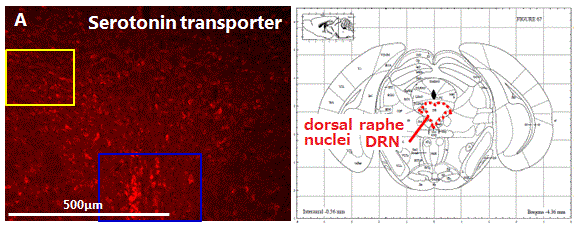
Experimental conditions
- Samples: Frozen sections of the dorsal raphe nuclei from the brain of 10-week-old ICR mice
- Staining method: Fluorescent antibody staining
- Activation condition: Microwave treatment in a citrate buffer (pH 6.0) for 10 minutes
- Antibody concentration: 1:100
- Primary antibody: This product
- Secondary antibody: Alexa 594 goat anti Rat IgG

Figs. B-D: Enlarged staining images of the area within a frame (in blue) in Fig. A
Figs. E-G: Enlarged staining images of the area within another frame (in yellow) in Fig. A
Image data are courtesy of Dr. Takuma and Hasebe of Graduate School of Dentistry, Osaka University.
Protocol example
- Wash the tissue section on the slide glass with phosphate-buffered saline containing 0.3 % Triton X-100® (PBST).
- Perform activation treatment in a citrate buffer (pH 6.0) with a microwave oven (10 minutes).
- Bring the tissue section back to ambient temperature.
- Wash the tissue section with 0.3 % PBST.
- Block the tissue section with 5% goat serum-PBS (1 hour).
- Incubate the tissue section with the primary antibody (this product) diluted 100-fold with 0.3 %PBST (4°C, overnight).
- Wash the tissue section with 0.3 % PBST.
- Incubate the tissue section with the secondary antibody (room temperature, 2 hours).
- After washing with 0.3% PBST, mount the tissue section.
Triton X-100 is a registered trademark of Dow Chemical Company.
Usage example – Immunohistochemical staining (DAB staining)
Fluorescence signals were noted in the neuronal cell body.
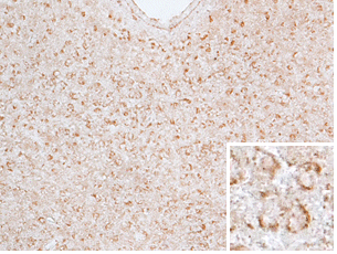 |
Experimental conditions
|
Image data are courtesy of Dr. Takuma and Hasebe, Graduate School of Dentistry, Osaka University in Japan.
Protocol example
- Wash the tissue section on the slide glass with phosphate-buffered saline containing 0.3 % Triton X-100® (PBST).
- Perform activation treatment in a citrate buffer (pH 6.0) with a microwave oven (10 minutes).
- Bring the tissue section back to ambient temperature.
- Wash the tissue section with 0.3 % PBST.
- Treat the tissue section with 40% methanol containing 0.3 % H2O2 (30 minutes)..
- Block the tissue section with 5% goat serum-PBS (1 hour).
- Incubate the tissue section with the primary antibody (this product) diluted 100-fold with 0.3 %PBST (4°C, overnight).
- Wash the tissue section with 0.3 % PBST.
- Incubate the tissue section with the biotinylated secondary antibody (room temperature, 2 hours).
- Wash the tissue section with 0.3 % PBST.
- Incubate the tissue section with DAB. After dehydration treatment, mount the tissue section.
Triton X-100 is a registered trademark of Dow Chemical Company.
Usage example – Flow cytometry
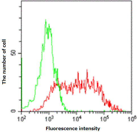
Green: 293T cells
Red: 293T cells expressing mouse serotonin transporter
Antibody concentration: 0.1 µg/mL
Product List
for Immunochemistry
- Manufacturer : FUJIFILM Wako Pure Chemical Corporation
- Storage Condition : Keep at -80 degrees C.
|
Comparison
|
Product Number
|
Package Size
|
Price
|
Inventory
|
|---|---|---|---|---|
|
50uL
|
|
Inquire |
for Immunochemistry
- Manufacturer : FUJIFILM Wako Pure Chemical Corporation
- Storage Condition : Keep at -20 degrees C.
|
Comparison
|
Product Number
|
Package Size
|
Price
|
Inventory
|
|---|---|---|---|---|
|
50uL
|
|
Inquire |
for Immunochemistry
- Manufacturer : FUJIFILM Wako Pure Chemical Corporation
- Storage Condition : Keep at -20 degrees C.
|
Comparison
|
Product Number
|
Package Size
|
Price
|
Inventory
|
|---|---|---|---|---|
|
10uL
|
Discontinued
|
||
|
50uL
|
Discontinued
|
Related Information
Category
- Cell Culture
- Nerve Cell Culture
- Antibody
- Life Science
- Neuroscience
- Antibody
Product content may differ from the actual image due to minor specification changes etc.
图片仅供参考,请以实物为准。
若本网站没有及时更新,请大家谅解!
正文中列出的所有试剂只能用于测试或研究,不能作为”药品”,”食品”,”家庭用品”等使用。
我司所销售的化学试剂、原料等所有产品(包括但不限于抗生素类、蛋白质类、试剂盒类产品等)仅限用于科学研究用途,不得作用于人体。
库存信息随时变更,准确货期以下单时确认为准。
重要提醒:该中文说明为国内翻译版本,仅供参考,与官网英文版有不符之处,以英文版说明为主。
