Wako 和光纯药 011-27991 Anti Iba1, Goat Iba1抗体,山羊源多克隆抗体说明书
【Background】
Iba1 is a protein highly expressed in microglia and macrophages with a molecular weight of about 16.7 kDa1). The protein is a commonly known microglial marker in the nervous system.This item is a goat polyclonal antibody that reacts with Iba1.
For Research Use Only. Not for use in diagnostic procedures or therapeutic use.
小胶质细胞标记
Iba1是一种约17 kDa的蛋白,在神经系统小胶质细胞中特异性表达,经常被用作小胶质细胞标记物。 本产品是识别Iba1的山羊多克隆抗体。
【Description】
[Purification] Purified from the goat serum by antigen-affinity chromatography
[Reactivity] Reacts with Iba1
[Antigen] Synthetic peptide corresponding to the C-terminus of Iba1
[Clone No.] -( polyclonal)
[Species cross reactivity] Mouse and Rat(Other species have not been tested)
[Host] Goat
[Concentration] indicated on the label
[Formulation] TBS
[纯化]通过抗原亲和层析从山羊血清中纯化
[反应性]与Iba1反应
[抗原]对应于Iba1 C末端的合成肽
[克隆号]-(多克隆)
[物种交叉反应]小鼠和大鼠(尚未测试其他物种)
[主持人]山羊
标签上指示的[浓度]
[配方] TBS
【Applications】
Immunohistochemistry( frozen section) 1 : 250-1,000
Immunohistochemistry( paraffin section) 1 : 250-1,000
Western Blot 1 : 1,000
Optimal concentration should be determined by each laboratory
for each application.
【Storage】
Store at -20℃ . Avoid repeated freeze and thaw.
【Package】
100μL
【Recommended protocol( Immunohistochemistry frozen section)】
Wistar rat or ICR mouse was perfusion-fixed with 4% paraformaldehyde.
replaced sucrose, and prepared 25μm brain section
by microtome.
Wash : 0.3% TritonX-100 in PBS, 5 min × 3
↓
Blocking : 1% BSA and 0.3% TritonX-100 in PBS, 2 hour, RT
↓
Primary antibody : goat anti-Iba1 (1/1000), 1% BSA, and 0.3%
TritonX-100 in PBS, overnight, 4℃
↓
Wash : 0.3% TritonX-100 in PBS, 5 min × 3
↓
Secondary antibody : AlexaFluor488 anti-goat IgG (1/1000,
Jackson Immuno Research Laboratories #705-545-147), 1%
BSA, and 0.3% TritonX-100 in PBS, 2 hour, RT
↓
Wash : 0.3% TritonX-100 in PBS, 5 min × 3
↓
Mount
【Reference】 参考文献
1) Imai, Y., Ibata, I., Ito, D., Ohsawa, K. and Kohsaka, S. : Biochem.
Biophys. Res. Commun., 224, 855( 1996).
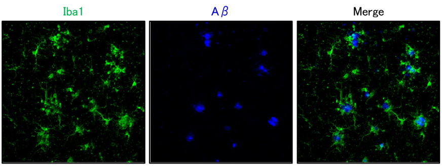
◆应用实例 1:免疫组织染色(荧光染色)
Immunohistochemistry (fluorescent staining)

数据提供:国立长寿医疗研究中心榊原老师
样品:阿尔茨海默病模型小鼠(APPNL-G-F 小鼠)大脑新皮层冰冻切片
一抗:抗Iba1,山羊多克隆抗体(1:1,000)
二抗:Alexa Fluor488标记抗山羊IgG
Aβ染色:0.001 % FSB溶液(淀粉样蛋白染色荧光探针)

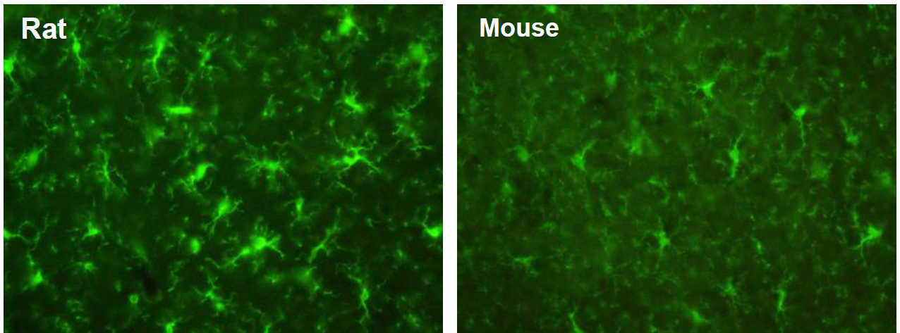
数据提供:创价大学理工学部中嶋老师
样本:大鼠(左)以及小鼠(右)大脑皮质冰冻切片
一抗:抗Iba1,山羊多克隆抗体(1:250)
二抗:Alexa Fluor488标记抗山羊IgG
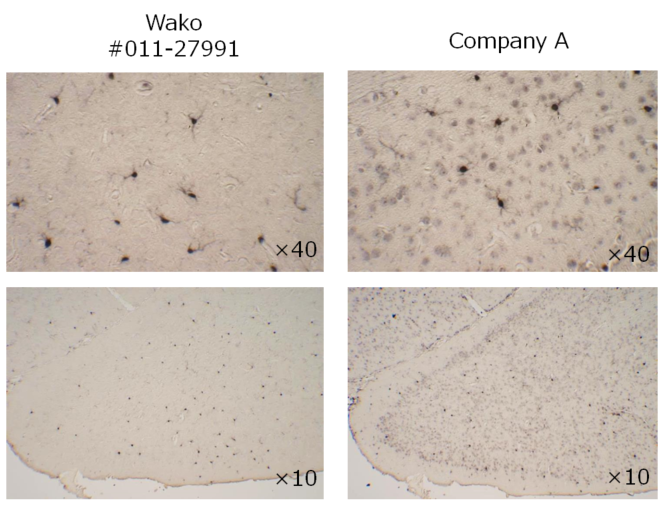
◆应用实例 2:免疫组织染色(DAB染色)

样品:小鼠脑额叶石蜡切片
一抗:抗Iba1,山羊(1:1,000)
二抗:抗山羊IgG,生物素标记
抗原激活:10 mM柠檬酸盐缓冲液(pH 6),90°C,处理10 min
◆应用实例3:蛋白印迹 Western Blotting
