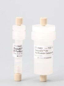
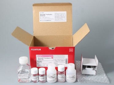
Wako MassivEV EV纯化分离柱 PS/MassivEV EV纯化缓冲液套装

Wako MassivEV EV纯化分离柱 PS/MassivEV EV纯化缓冲液套装

用于大规模纯化外泌体等细胞外囊泡的分离柱

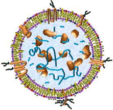
以外泌体为代表的细胞外囊泡(EV: Extracellular Vesicles)是由细胞释放的脂质双分子层微囊泡,内含核酸、蛋白和脂质等。作为生物体内细胞间的通信媒介,外泌体相关的研究成为免疫、癌症、神经和代谢等多个领域的研究热点。

EV的应用领域广泛,有望应用于治疗药物、生物标志物和药物递送系统 (DDS)。尤其是间充质干细胞(MSC: Mesenchymal stem cell)分泌的EV具有抗炎、抗纤维化和组织修复等作用1),除医疗领域外,在化妆品和食品等领域也备受关注。

EV的实际应用需要能够高效且大规模分离和纯化高纯度EV的技术。富士胶片和光采用与金泽大学医学系免疫学的华山教授共同开发的分离和纯化技术“PS亲和法”2),开发了用于大规模纯化EV的MassivEV™ EV Purification Column PS(MassivEV™ 细胞外囊泡(EV)纯化分离柱 PS)。配合使用专用的缓冲液MassivEV™ Purification Buffer Set(MassivEV™ 细胞外囊泡(EV)纯化缓冲液套装),可从支持规模放大的细胞培养上清中简易分离和纯化EV。

图1.jpg 图2.jpg
MassivEV™ EV Purification Column PS MassivEV™ Purification Buffer Set

◆产品概要

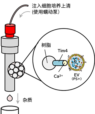
MassivEV™ EV Purification Column PS和MassivEV™ Purification Buffer Set是用于从大规格细胞培养上清中分离和纯化外泌体等细胞外囊泡(EV)的分离柱和专用缓冲液。采用与EV膜表面的磷酯酰丝氨酸(PS)钙离子依赖性结合Tim4的“PS亲和法”*,可简易纯化高纯度EV。

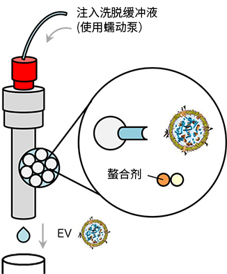
*利用与EV膜成分的磷酯酰丝氨酸(PS)特异性结合Tim4((T-cell immunoglobulin domain and mucin domain-containing protein 4))的纯化方法。即利用与PS钙离子依赖性结合Tim4的性质,在钙离子存在下,固定在载体上的Tim4可捕获样品中的EV。通过清洗去除杂质后,加入EV Elution Buffer(螯合剂),低损伤状态下从载体上的Tim4的洗脱和纯化EV的方法。

◆特点

● 可从大规格(10 mL至公升级别规格)细胞培养上清中高效分离和纯化高纯度EV

● 无需切向流过滤(TFF)系统等昂贵设备 ※只需一个蠕动泵

表1 从200 mL间充质干细胞培养上清中分离和纯化EV的对比(富士胶片和光评估)

  MassivEV™ TFF+ AEX TFF+SEC
可纯化的EV   PS阳性EV   取决于样品   取决于样品
纯度   高   低   低
纯化步骤数   1 个步骤

– PS 亲和法

  2 个步骤

– TFF 系统

– 阴离子交换色谱法

  2 个步骤

– TFF 系统

– 尺寸排阻色谱法

EV粒子产量(参考值)   1.7×10¹¹ particles   1.1×10¹¹ particles   0.7×10¹¹ particles
纯化所需时间   8 h   10 h   10 h

※AEX: 阴离子交换色谱法; SEC:尺寸排阻色谱法

<参考> 一只小鼠的EV建议给药量3-5):1.0×10⁹ particles/mouse

适应性

细胞培养上清(MSC等):10 mL至公升级别规格

※ 从10 mL以下的细胞培养上清中分离EV时,请使用MagCapture™ Exosome Isolation Kit PS Ver.2(产品编号:290-84103

处理样品量

  1 mL (产品编号:131-19491) 5 mL (产品编号:137-19493)
样品量※1 200 mL 1 L
动态结合能力※2 5×10¹¹ particles/mL 树脂 2.5×10¹² particles/5 mL树脂

※1 MSC细胞培养上清中一次纯化可处理的大概样品量。样品处理量因细胞培养上清中所含的EV粒子数而异。处理同一样品时,可重复使用分离柱,已确认最多可使用五次(初次使用1次、重复使用4次)。

※2 使用MSC来源EV的研究结果。可能因细胞种类等条件而异。

原理

PS亲和法利用与EV膜表面的磷酯酰丝氨酸(PS)特异性结合的Tim4,是富士胶片和光开发的EV分离方法。

PS-Tim4结合的高特异性配合螯合剂的温和洗脱,可使EV在完整状态下高纯度分离。


结合

3017065792968548.jpg

洗脱

3017065793479066.jpg

图1 MassivEV™ EV Purification Column PS(PS亲和法)分离和纯化细胞外囊泡的原理

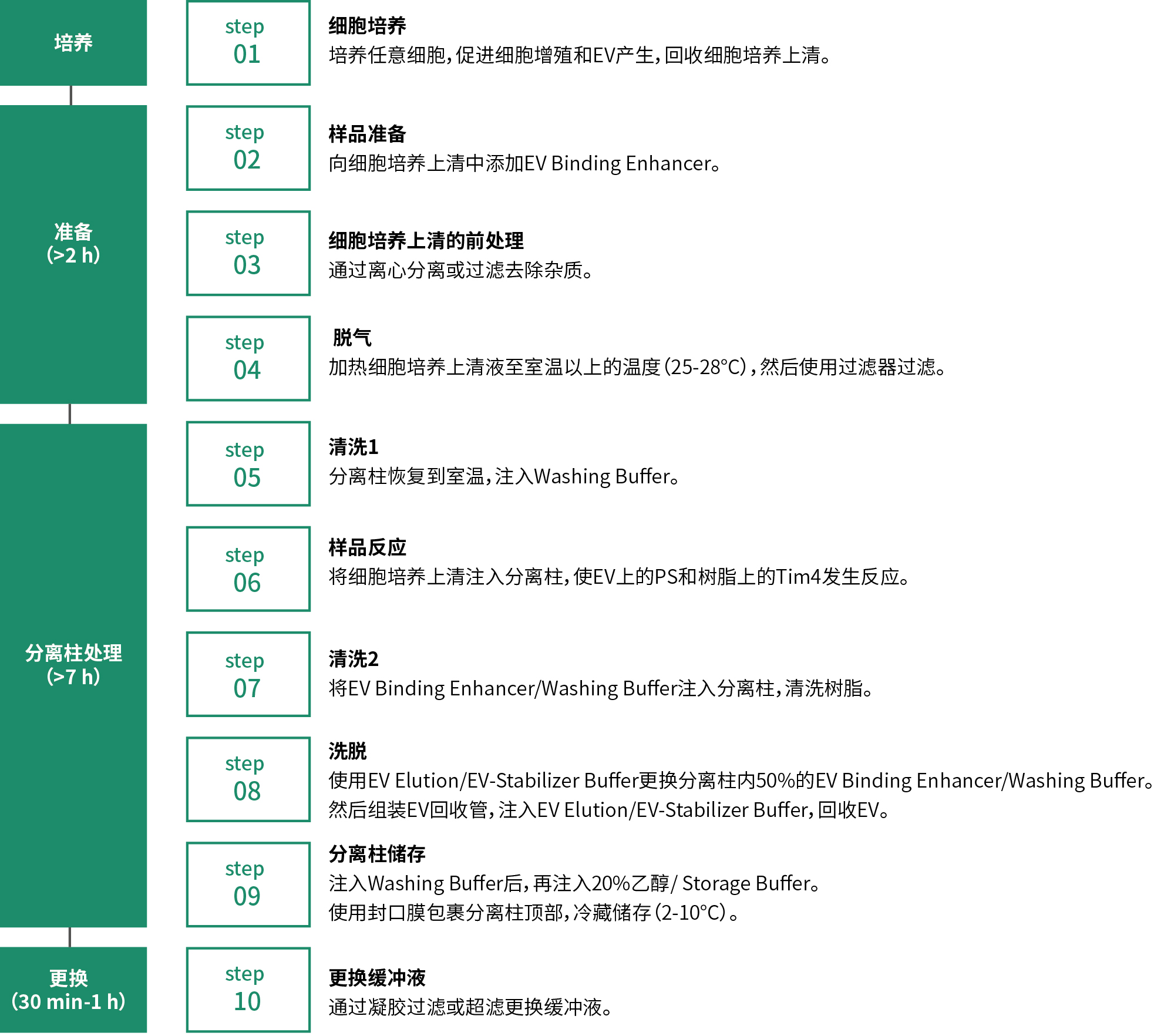
◆实验方案

2617062561475619.jpg

◆数据

性能数据

与传统方法对比

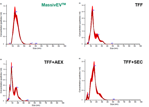
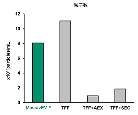
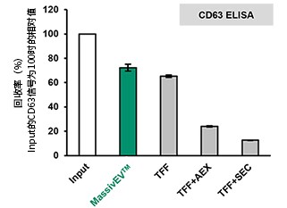
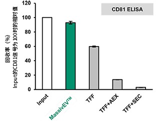
在增殖培养基(MSCulture™/ 10% FBS)及EV生产培养基(EV-Up™)中培养骨髓来源的MSC,回收细胞培养上清后,使用0.22 μm过滤器过滤。取200 mL经过滤处理的细胞培养上清用作样品,采用以下4种方法进行分离和纯化。纯化后的EV溶液分别使用Nanoparticle Tracking Analysis (NTA)和ELISA进行分析。

<分离方法>

MassivEV™   MassivEV™ EV Purification Column PS / MassivEV™ Purification Buffer Set (本产品, PS亲和法)

TFF                仅切向流过滤(500 kDa)

TFF+AEX      切向流过滤(500 kDa)+阴离子交换色谱法

TFF+SEC       切向流过滤(500 kDa)+尺寸排阻色谱法

(1)使用NTA 进行粒子分析


2617062568663115.png 2617062569104694.png

【结果】

与TFF相比,MassivEV™ 纯化得到的粒子数量少,但与 TFF+AEX 或 TFF+SEC 相比,MassivEV™ 纯化得到的粒子数量更多

(2)使用CD63 ELISA和CD81 ELISA的EV回收率比较(n=3)


CD63.jpg CD81.jpg

【结果】

相比传统方法,使用MassivEV™ 的EV回收率更高。


产品列表
产品编号 产品名称 产品规格 产品等级 备注
131-19491 MassivEV™ EV Purification Column PS
MassivEV™ 细胞外囊泡(EV)纯化分离柱 PS
1 mL 基因研究用
137-19493 MassivEV™ EV Purification Column PS
MassivEV™ 细胞外囊泡(EV)纯化分离柱 PS
5 mL 基因研究用
295-96601 MassivEV™ Purification Buffer Set
MassivEV™ 细胞外囊泡(EV)纯化缓冲液套装
1 mL×10次用
(5 mL×2次用)
基因研究用
以上MassivEV™ EV Purification Column PS、MassivEV™ Purification Buffer Set请配合一同使用

相关产品
间充质干细胞(MSC)增殖用培养基
产品编号 产品名称 产品规格 产品等级 备注
132-19345 MSCulture™ High Growth Basal Medium
MSCulture™ High Growth基础培养基
500 mL 细胞培养用
133-19331 MSCulture™ High Growth Supplement
MSCulture™ High Growth添加剂
5 mL 细胞培养用
以上MSCulture™ High Growth基础培养基、MSCulture™ High Growth添加剂请配合一同使用
间充质干细胞(MSC)EV生产用培养基
产品编号 产品名称 产品规格 产品等级 备注
053-09451 EV-Up™ EV Production Basal Medium for MSC, AF
EV-Up™ 间充质干细胞外泌体生产用基础培养基,无动物源成分
95 mL 细胞培养用
298-84001 EV-Up™ MSC EV Production Supplement, AF
EV-Up™ 间充质干细胞外泌体生产用添加剂,无动物源成分
100 mL用 细胞培养用
以上EV-Up™基础培养基、EV-Up™添加剂请配合一同使用
外泌体分离和纯化试剂盒
产品编号 产品名称 产品规格 产品等级 备注
294-84101 MagCapture™ Exosome Isolation Kit PS Ver.2
MagCapture™外泌体提取试剂盒PS Ver.2
2 tests 基因研究用
290-84103 MagCapture™ Exosome Isolation Kit PS Ver.2
MagCapture™ 外泌体提取试剂盒PS Ver.2
10 tests 基因研究用
适用于从10 mL以下的细胞培养上清和体液样品中分离和纯化EV

FAQ About product usage

What reagents, instruments, and equipment are required for the EV purification using this column?
The reagents, instruments, and equipment required for the use of this product are listed below.

-Reagents-

  • MassivEV™ Purification Buffer Set (Product Number: 295-96601)
  • Cell culture medium
  • EV production medium (Product Number: 053-09451 / 298-84001)
  • 100% Ethanol
  • Ultrapure water

-Instruments and Equipment-
*The following instruments may or may not be required depending on the operation. For more details, please see instruction manual.

  • Centrifuge
  • Cell strainer(e.g. FALCON, Product Number: 352340)
  • 5 μm filter(e.g. GVS, Product Number: 1215396)
  • 0.8 μm filter(e.g. GVS, Product Number: 1214568)
  • 0.22 μm filter(e.g. Corning, Product Number: 431118)
  • Incubation or water bath (which can be warmed at 37℃)
  • Peristaltic pump (e.g. Repligen, Product Number: ACJR-U10-R)
  • Fluid transfer tubing (see support document)
  • Collection tube (e.g. Corning, Product Number: 430791)
  • Ultrafiltration membrane [100kDa/PES material] (e.g. Sartorius, Product Number: VS0141)
  • Gel-filtration columns(e.g. Cytiva, Product Number: 28918004)
Do you have any recommendations for tubing suitable for liquid transfer? How should this tubing be connected?
We don’t have specific recommendations, provided the tubing can control the flow rate and withstand pressure. Ensure the tubing is compatible with your peristaltic pump.
For guidance on connecting the tubing, please refer the support document titled:
Example of a purification system using the MassivEV EV Purification Column PS
How should I choose between centrifugation and filtration for the pretreatment of cell culture supernatants?
Your choice between centrifugation and filtration will depend on the facilities, instruments, and equipment your laboratory already has. The decision does not need to be influenced by the sample type or its specific application.
For buffer exchange, how should I choose between gel filtration and ultrafiltration?
We recommend gel filtration when the sample is for administration to animals.
What volume of the EV solution is obtained during elution?
The volume is 4 mL for a 1 mL column, and 20 mL for a 5 mL column.
How long can a column wrapped in Parafilm be stored?
Based on our experience at Fujifilm Wako, a column can be stored for approximately 1.5 months.
Can the column be used multiple times?
The column can be reused for the same sample up to 5 times, including the initial use and four subsequent reuses.
For each reuse, begin with Cleaning 1, as detailed in the “Procedures” section (5) of the instruction manual.

Troubleshooting

How should air that has entered the column be removed?
To remove air from the column, try passing 20% or 70% ethanol through it at the flow rates specified below. Note that prolonged or repeated exposure to air can degrade the resin’s performance. For more information, consult the instruction manual.

Flow Rates
– 1 mL column: 0.85 mL/min
– 5 mL column: 4.25 mL/min

About commercial or for-profit purposes

Can I use this product for commercial or for-profit purposes?
Please use this product for research purposes only. If you wish to use it for commercial or for-profit purposes,

Alias

  • MassivEV™ Exosome Purification Column PS

For research use or further manufacturing use only. Not for use in diagnostic procedures.

Product content may differ from the actual image due to minor specification changes etc.

If the revision of product standards and packaging standards has been made, there is a case where the actual product specifications and images are different.

The prices are list prices in Japan.Please contact your local distributor for your retail price in your region.

免责声明
1. 本公司密切关注本网站发布的内容,但不保证发布内容的准确性、完整性、可靠性和最新性等。2. 本公司不保证使用本网站期间不会出现故障或计算机病毒污染的风险。3. 无论何种原因,使用本网站时给用户或第三方造成的任何不利或损害,本公司概不负责。此外,对于用户与其他用户或第三方之间因本网站发生的任何交易、通讯或纠纷,本公司概不负责。

4. 本网站可提供的所有产品和服务均不得用于人体或动物的临床诊断或治疗,仅可用于科研等非医疗目的。如任何用户将本网站提供的产品和服务用于临床诊断或治 疗,以及其他特定的用途或行为,本公司概不保证其安全性和有效性,并且不负任何相关的法律责任。


Wako和光纯药-Aluminium Potassium Sulfate

Wako和光纯药-Aluminium Potassium Sulfate

Aluminium Potassium Sulfate

品牌:Wako
CAS No.:10043-67-1
储存条件:室温

Wako
和光纯药
011-02035


Aluminium Potassium Sulfate 500 g 10043-67-1

* 干冰运输、大包装及大批量的产品需酌情添加运输费用

* 零售价、促销产品折扣、运输费用、库存情况、产品及包装规格可能因各种原因有所变动,恕不另行通知,确切详情请联系上海金畔生物科技有限公司。

请联系客服
若本网站没有及时更新,请大家谅解!
正文中列出的所有试剂只能用于测试或研究,不能作为”药品”,”食品”,”家庭用品”等使用。
我司所销售的化学试剂、原料等所有产品(包括但不限于抗生素类、蛋白质类、试剂盒类产品等)仅限用于科学研究用途,不得作用于人体。

Wako和光纯药 L-丙氨酸 L-Alanine

Wako和光纯药 L-丙氨酸 L-Alanine

品牌:Wako
CAS No.:56-41-7
储存条件:室温

产品编号 等级 规格
010-01042 JIS Special Grade 25 g

* 干冰运输、大包装及大批量的产品需酌情添加运输费用

* 零售价、促销产品折扣、运输费用、库存情况、产品及包装规格可能因各种原因有所变动,恕不另行通知,确切详情请联系上海金畔生物科技有限公司。

请联系客服 若本网站没有及时更新,请大家谅解!
正文中列出的所有试剂只能用于测试或研究,不能作为”药品”,”食品”,”家庭用品”等使用。
我司所销售的化学试剂、原料等所有产品(包括但不限于抗生素类、蛋白质类、试剂盒类产品等)仅限用于科学研究用途,不得作用于人体。

Wako Alumina, Activated 活性氧化铝

Wako Alumina, Activated 活性氧化铝

品牌:Wako
CAS No.:1344-28-1
储存条件:室温

产品编号 等级 规格
010-01525 for Column Chromatography 500 g

* 干冰运输、大包装及大批量的产品需酌情添加运输费用

* 零售价、促销产品折扣、运输费用、库存情况、产品及包装规格可能因各种原因有所变动,恕不另行通知,确切详情请联系上海金畔生物科技有限公司。

请联系客服 若本网站没有及时更新,请大家谅解!
正文中列出的所有试剂只能用于测试或研究,不能作为”药品”,”食品”,”家庭用品”等使用。
我司所销售的化学试剂、原料等所有产品(包括但不限于抗生素类、蛋白质类、试剂盒类产品等)仅限用于科学研究用途,不得作用于人体。

Wako和光纯药297-51501 SLP Reagent Set 3mL 替代品:296-81001

Wako和光纯药297-51501 SLP Reagent Set 3mL 替代品:296-81001

SLP Reagent Set (真菌和细菌感染快速检测)
——肽聚糖和(1→3)-β-D-葡聚糖定量检测
家蚕的淋巴有一种自我保护机制,被称为“酚氧化酶原串联反应”。当受外界入侵(如细菌和真菌)时,
这个串联反应参与黑色素的形成并使机体免受攻击。这个反应由细菌中的肽聚糖或是真菌、酵母中的(1→
3)-β-D-葡聚糖引发,该反应中的酚氧化酶原被激活。也有人认为丝氨酸蛋白酶也参与了该反应,但是没
有从这方面阐述的资料。
已消毒的SLP包括了所有参与该反应的因子,再生后的溶液中含有L-3,4-苯丙氨酸(L-3,4-
dihydroxyphenylalanine, DOPA),该物质可以作为肽聚糖或β-葡聚糖所激发的酚氧化酶的底物,DOPA被
酚氧化酶氧化,形成黑色素。肽聚糖是绝大多数细菌细胞壁的组成成分,而β-葡聚糖广泛的分布于真菌
的细胞壁中。因此,SLP通过测定黑色素的形成情况,进而定性或定量检测细菌和真菌的污染。
特点
1. 用于肽聚糖和(1→3)-β-D-葡聚糖的定量检测,检测敏感。
2. 使用酶标仪或内毒素检测仪,能对肽葡聚糖和β-葡聚糖精确定量。
3. 不需任何特殊器械即可通过肉眼检测出细菌和真菌的污染。
4. 冻干SLP的在2-10℃至少可保存18个月。
原理见:http://www.cxbio.com/news/news_details.asp?id=9588
当使用酶标仪或内毒素检测仪时,通过测定反应的始动时间和由颜色变化而导致的吸光度的变化,即可对
肽聚糖或β-葡聚糖进行定量。
所含试剂(所有试剂均存放于2-10℃):
1. 冻干SLP             for 3ml
使用酶标仪或者目测约50Test,使用内毒素检测仪约30Test
2. 冻干底物             for 3ml
含有作为酚氧化酶底物的DOPA
3. 稀释液           4ml
含有good’s buffer
试剂准备:
(A) 底物中加入3ml稀释液,轻轻摇晃至底物完全溶解,注意避免碰到橡胶帽。
(B) 所有底物溶液加入冻干的SLP中,轻轻摇晃至SLP完全溶解,同样,注意避免碰到橡胶帽。此溶液即为
SLP检测液。
操作步骤
1.  使用酶标板(微孔板)目测
(A) 分别稀释肽聚糖和β-葡聚糖参照物,做两个不同浓度的参照系列,每孔加入0.05ml的参照物稀释液
或者待测样品
(B) 每孔加入0.05mlSLP检测液
(C) 温和摇晃后,酶标板于30℃孵育一小时
(D) 肉眼判断:反应液中有黑色素生成的为阳性,其它的为阴性。
(E) 参考物的最低浓度(即能生成黑色素的最小浓度)不应在SLP检测液的最低检测浓度以下。
(F) 样品中SLP反应物浓度是否在SLP检测液浓度的最低检测范围内,决定了样品是阳性或阴性。
2. 使用酶标仪检测
(A) 样品和参考物的稀释系列各0.05ml分别加入酶标板孔中
(B) 迅速在每孔中加入0.05mL SLP检测液。好用消毒过的加液器。
(C) 在以下条件下使用酶标仪上测定光吸收值:
检测波长:  650nm
检测模式:  动态检测
反应时间:  90分钟
检测温度:  30℃
自动混匀:  一次
(D) 绘制双对数回归(regression of Log-Log)标准曲线,横轴为SLP参考物的浓度,坚轴为反应时间
(E) 通过标准曲线上的反应始动时间,计算SLP反应物质的浓度。
3. 使用内毒素检测仪做SLP检测,其操作步骤可以按照仪器生产商提供的操作手册中所述的细菌内毒素检
测方法操作。内毒素检测仪可能会发出反应混合液缺乏透射光的信号。忽视这个的警告,测定结果不会受
此影响。
关联试剂:
297-51501   SLP Reagent Set    (定量的需要另配以下标准品使用)
162-18101   Peptidoglycan, from Micrococcus luteus肽聚糖    2ml
030-09903   Curdlan  (1→3)-β-D-葡聚糖    1g
注意事项
1. 器械应250℃干热灭菌至少2小时,并避免被SLP反应物污染
2. SLP冻干粉溶解后,其溶液应存于0-4℃,并尽早使用
3. 注意在器械储存期间避免其被SLP反应物质污染
4. SLP检测液的敏感性很大程度上受离子强度的影响
5. 该试剂盒仅用于研究,不可用于诊断;仅用于体外,不可用于人或动物体内。
(本资料只供参考,以原包装说明书为准)
常见使用说明:
1.      因为SLP是被PG(肽聚糖)或BG[(1→3)-β-D-葡聚糖]所引发,在操作和使用本试剂时,单一灭
菌器皿或者使用无热源器皿都不够的,必须还要同时无PG和BG;
2.      如果是用玻璃器皿,清洗干净后,需要在250度下干热灭菌2小时;
3.      如果是一次性无菌器皿,在使用之前必须进行测试能否适用于此项检测(即注1);
4.      如果使用过后还有剩余,我们认为其很可能被样品污染,不建议继续使用。以前我们也遇到类似
的问题,但所有的都是因为污染造成。如果用户一定要保留一些SLP溶液,建议在未使用前进行无菌分装
并0-4℃保存;好是将SLP冻干粉在无菌无污染条件下,按照使用计划量预先分装后0-4℃保存。
其他相关产品

297-51501 SLP Reagent Set  3mL用

293-58301 SLP-HS Single Reagent Set  20回用

198-01055 Sodium Acetate Trihydrate  500g

194-13321 Sodium Alginate 80-120  100g

198-01675 Sodium Chloride  500g

199-08361 Sodium Dextran Sulfate 5,000

194-13402 Sodium Dextran Sulfate 5000  25g

197-09705 Sodium Dihydrogenphosphate  500g

192-02815 Sodium Dihydrogenphosphate Dihydrate  500g

190-02811 Sodium Dihydrogenphosphate Dihydrate  100g

192-07935 Sodium Hydroxide  500g

197-03345 Sodium Sulfate  500g

197-07125 Sodium Sulfate  500g

191-03985 Soluble Starch 和光一級  500g

199-16291 Stevioside Standard  100mg

196-00015 Sucrose  500g

191-04502 Sulfanilamide  25g

204-08343 Testosterone  5g

203-00432 Tetraethylammonium Bromide  25g

205-00431 Tetraethylammonium Bromide  100g

208-16771 TFNG Standard  20mg

124-02275 Trilithium Citrate Tetrahydrate  500g

206-15591 Trinexapac Standard  200mg

190-05535 Trisodium Citrate Dihydrate  500g

208-09223 Trypsin Inhibitor, from Soybean  1g

028-03386 t-Butyl Alcohol 500ml

223-02112 VA-044  25G

226-01306 Vancomycin Hydrochloride  5g

302-07111 Vitamin E Reference Standard  1kit

234-01565 WakogelR 50C18  500g

396-01731 XLD 日局試験用  300g

035-17981 α-Carotene Standard  10mg

354-03232 γ-Phenylpropyl Acetate  25g

242-00641 Xylobiose  100mg

334-30183 Z-Glu-Tyr  1g

263-00271 Zinc Chloride  10g

033-08332 α-Cyclodextrin  25g

034-13632 Chitin  25g

030-09903 Curdlan  1g

342-01875 EDTA-3NA  500g

030-05525 Citric Acid  500g

192-09071 Silicon Dioxide, 99.9%  100g

192-06031 Silicon Standard Solution  100ml

194-00832 Silver Nitrate  25g

196-12781 Simeconazole Standar  200mg

Wako新型PS亲和法Exosome外泌体纯化试剂盒

Wako新型PS亲和法Exosome外泌体纯化试剂盒

外泌体和其他细胞外囊泡(EVs)属于小细胞膜囊泡,包含蛋白,mRNA,microRNA,DNA和脂类,是由不同细胞分泌,并且在体液包括血液、唾液、 尿液、脑脊髓液(CSF)和乳液中稳定存在。这些EVs被当做细胞和细胞沟通的信使,不同疾病的生物标志物。提取外泌体的常规方法有:超离,使用表面抗原 的抗体进行亲和层析,和用多聚物试剂进行沉淀。然而,这些方法在回收效率,纯度和可操作性上不是非常令人满意。

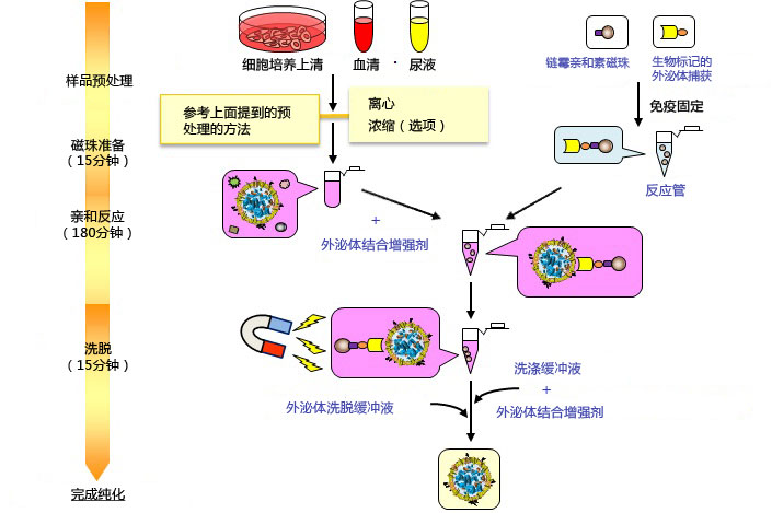
MagCapture™Exosome Isolation Kit PS采用磁珠和磷脂酰丝氨酸(PS)结合蛋白(PS亲和法)的方法进行纯化。该方法可以很方便获得高纯度的外泌体和其他来自细胞培养基和体液的EVs(通 过正常的微滤方法获得高产量)。如果想要获得更高纯度的外泌体,请使用10,000×g获得上清。该试剂盒利用中性pH的金属螯合试剂,将捕获的EVs从 磁珠上洗脱下来,可以获得完整的外泌体和其他EVs。纯化的完整外泌体和其他EVs可用于不同实验,包括电镜分析,纳米粒子追踪分析,EVs的操作和分析。

细胞膜表面磷脂酰丝氨酸的亲和方法

anf[1]-cn.jpg

使用磷脂酰丝氨酸(PS)结合蛋白,细胞外囊泡在金属离子依赖方式捕获,接着用金属粒子螯合试剂进行洗脱。

◆优点・特色

nezumi-1[1]-cn.jpg


  使用新型亲和纯化方法

  ● 通过PS亲和分子回收

● 低背景值

● 在中性条件下通过螯合试剂进行温和洗脱

※ 可获得高纯度,完整的外泌体

捕获.JPG


无需进行超离

● 使用磁珠改进了操作

● 流程优化

※ 高重复性

与其他纯化方法相比

2014532734317504.gif

方法 纯度 外泌体回收率 获得囊泡的完整性
MagCapture™

(PtdSer亲和方法)

■■■
超离 ■■
多聚物沉淀 × ■■■■
外泌体表面抗原亲和方法(使用抗体) ×

样品类型:细胞培养上清,血清,血浆,尿液等。

  ◆产品列表

产品名称 包装规格 和光编号 保存条件
MagCapture™
Exosome Isolation Kit PS
10次 293-77601 保存在2-10°C
试剂盒成分

(1) 链霉亲和素磁珠
(2)生物素标记的外泌体捕获
(3)外泌体捕获免疫固定缓冲液
(4) 外泌体结合增强剂(x 500)
(5) 洗涤缓冲液
(6) 外泌体洗脱缓冲液
(7) 反应管

<10 次>
600 μL x 1 管
100 μL x 1 管
35 mL x 1 瓶
500 μL x 1 管
75 mL x 2 瓶
5 mL x 1 瓶
22 管

*每个test里的磁珠可以循环使用共5次,即试剂盒的实际使用量为:10tests*5次/Kit

产品编号 产品名称 产品规格
293-77601 MagCapture™ Exosome Isolation Kit PS
MagCapture™ 外泌体提取试剂盒PS
10次用
299-77603 MagCapture™ Exosome Isolation Kit PS
MagCapture外泌体提取试剂盒PS
2次用
297-79201 PS Capture™ Exosome ELISA Kit (Anti Mouse IgG POD)
PS Capture™外泌体ELISA试剂盒(抗小鼠IgG POD)


品牌 产品编号 产品名称 等级 规格
Wako 299-77603 MagCapture™ Exosome Isolation Kit PS MagCapture™外泌体提取试剂盒PS for Genetic Research 2 tests
Wako 293-77601 MagCapture™ Exosome Isolation Kit PS MagCapture™外泌体提取试剂盒PS 10 tests
Wako 290-84103 MagCapture™ Exosome Isolation Kit PS Ver.2 MagCapture™外泌体提取试剂盒PS Ver.2 for Genetic Research 10 tests
Wako 294-84101 MagCapture™ Exosome Isolation Kit PS Ver.2 MagCapture™外泌体提取试剂盒PS Ver.2 for Genetic Research 2 tests
Wako 297-79201 PS Capture™ Exosome ELISA Kit (Anti Mouse IgG POD) PS Capture™外泌体ELISA试剂盒(抗小鼠IgG POD) 96 tests
Wako 297-79701 PS Capture™ Exosome Flow Cytometry Kit PS Capture™外泌体流式试剂盒 for Genetic Research 300 tests
Wako 298-80601 PS Capture™ Exosome ELISA Kit (Streptavidin HRP) PS Capture™外泌体ELISA Kit (链霉亲和素HRP) for Genetic Research 96 tests
Wako 290-80301 PS Capture™ Exosome Isolation Resin Kit PS Capture™外泌体提取树脂试剂盒 for Genetic Research 1 kit(0.5 mL浆状)
Wako 052-09301 Exosomes, from COLO201 cells, purified COLO201来源外泌体(已纯化) for Genetic Research 50 μL
Wako 292-83801 CD81-Capture Human Exosome ELISA Kit (Streptavidin HRP) CD81-Capture 人外泌体ELISA试剂盒(链霉亲和素HRP) for Genetic Research 96 tests

组织透明化原理

细胞膜表面磷脂酰丝氨酸的亲和方法

外泌体提取试剂盒

使用磷脂酰丝氨酸(PS)结合蛋白,细胞外囊泡在金属离子依赖方式捕获,接着用金属粒子螯合试剂进行洗脱

外泌体提取试剂盒的优点/特色

使用新型亲和纯化方法!

可获高纯度,完整的外泌体!

无需进行超离 !

组织透明化检测流程

产品编号 产品名称 产品规格 零售价
293-77601 MagCaptureTM Exosome Isolation Kit PS
Exosome外泌体纯化试剂盒
10次 8980

*每套试剂盒能完成5次回收实验,即实际使用量为10×5次/Kit

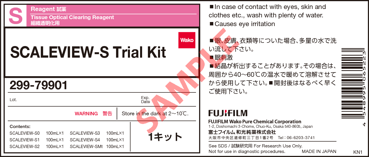
Wako 299-79901 组织透明化试剂盒 SCALEVIEW-S Trial Kit

Wako 299-79901 组织透明化试剂盒 SCALEVIEW-S Trial Kit

The original recipe reported by the Miyawaki team in 2011 termed Scale was an aqueous solution based on urea that is limited because the transparency process itself can damage the structures under study.
The research team spent 5 years improving the effectiveness of the original recipe to overcome this critical challenge, and the result is ScaleS, (we called SCALEVIEW-S) a new technique with many practical applications. SCALEVIEW-S creates transparent brains with minimal tissue damage, that can handle both florescent and immunohistochemical labeling techniques, and is even effective in older animals.
The new technique creates transparent brain samples that can be stored in SCALEVIEW-S solution for more than a year without damage. Internal structures maintain their original shape and brains are firm enough to permit the micron-thick slicing necessary for more detailed analyses.

Miyawaki 团队在 2011 年报告的原始配方称为 Scale 是一种基于尿素的水溶液,它是有限的,因为透明过程本身会损坏正在研究的结构。
研究团队花了 5 年时间改进原始配方的有效性以克服这一关键挑战,结果是 ScaleS,(我们称为 SCALEVIEW-S)一种具有许多实际应用的新技术。 SCALEVIEW-S 创造了组织损伤最小的透明大脑,可以处理荧光和免疫组织化学标记技术,甚至对老年动物也有效。
新技术创造了透明的大脑样本,可以在 SCALEVIEW-S 解决方案中存储一年多而不会损坏。 内部结构保持其原始形状,大脑足够坚固,可以进行更详细分析所需的微米厚切片。

Overview / Applications

Outline This product is for research use only. Do not administer it to human.
SCALEVIEW-S series are reagent used for ScaleS. ScaleS is tissue clearing method developed by Dr. Atsushi Miyawaki etc. This method uses solutions with mainly urea.
The tissue is made transparent and adjusted RI for microscopic observation by soaking in 6 kinds of solutions (SCALEVIEW-S0, SCALEVIEW-S1, SCALEVIEW-S2, SCALEVIEW-S3, SCALEVIEW-S4, SCALEVIEW-SMt). 2mm section can be made transparent in 1 day using the reagents.
The method can make a transparent tissue keeping the structure of proteins. Therefore it is possible to observe the tissue expressing fluorescent proteins, and to label by antibody and/or fluorescent dye after tissue clearing. It’s also applicable to not only mouse and rat brain but also human after dye. The transparent tissue can be observed for electric microscope or confocal microscope.
ScaleS is higher transparency than ScaleA2.

See the following website for more details.
* The cleaning solution, deScale Solution (041-34425) , is sold separately.

概述 / 应用

本产品仅供研究使用。不要将其施用于人类。
SCALEVIEW-S 系列是用于 ScaleS 的试剂。 ScaleS 是宫胁敦博士等开发的组织清除方法。该方法使用以尿素为主的溶液。
通过浸泡在 6 种溶液(SCALEVIEW-S0、SCALEVIEW-S1、SCALEVIEW-S2、SCALEVIEW-S3、SCALEVIEW-S4、SCALEVIEW-SMt)中使组织透明并调整 RI 用于显微观察。 2mm 切片可以在 1 天内使用试剂变透明。
该方法可以制成保持蛋白质结构的透明组织。因此可以观察表达荧光蛋白的组织,并在组织清除后用抗体和/或荧光染料进行标记。它不仅适用于小鼠和大鼠的大脑,也适用于染色后的人类。透明组织可用电镜或共聚焦显微镜观察。
ScaleS 的透明度高于 ScaleA2。

有关详细信息,请参阅以下网站。

* 清洁液 deScale Solution (041-34425) 单独出售。

Product List

Product Name
SCALEVIEW-A2 Tissue Optical Clearing Reagent
SCALEVIEW-S0 Tissue Optical Clearing Reagent
SCALEVIEW-S1 Tissue Optical Clearing Reagent
SCALEVIEW-S2 Tissue Optical Clearing Reagent
SCALEVIEW-S3 Tissue Optical Clearing Reagent
SCALEVIEW-S4 Tissue Optical Clearing Reagent
SCALEVIEW-SMt Tissue Optical Clearing Reagent
deScale Solution Tissue Optical Clearing Reagent

References  参考文献

  1. Hama,H.et al. : Nature Neuroscience 14, 1481(2011).
  2. Hama,H.et al. : Nature Neuroscience 18, 1518(2015).
  3. Hama H, et al. : Protocol Exchange (2016), doi:10.1038/protex.2016.019
  4. Molly E, Boutin, et al :Scientific Reports, 8, 11135 (2018).

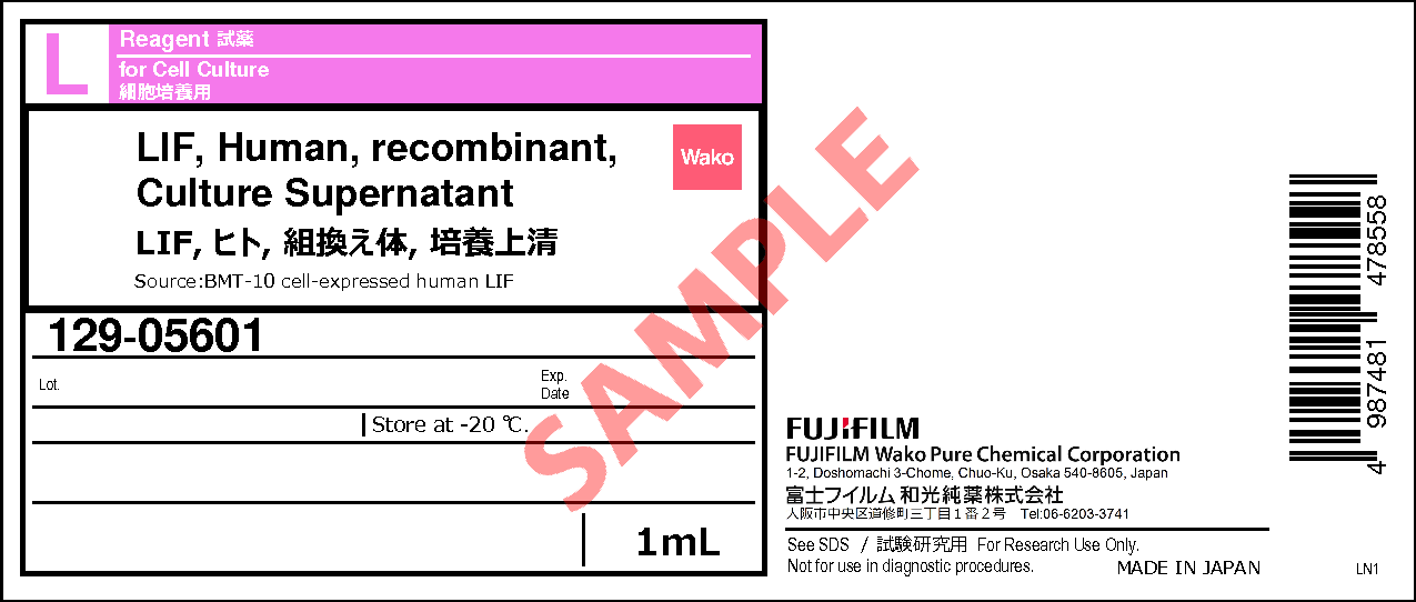
Wako 125-05603 重组人白血病抑制因子,细胞上清

Wako 125-05603 重组人白血病抑制因子,细胞上清

品牌:Wako
CAS No.:
储存条件:
-20℃

for Cell Culture

Manufacturer :  FUJIFILM Wako Pure Chemical Corporation

Storage Condition :  Keep at -20 degrees C.

129-05601 1mL
125-05603  1mL x10

Wako 290-80801 CUBIC Trial Kit CUBIC试剂盒

Wako 290-80801 CUBIC Trial Kit  CUBIC试剂盒

CUBIC Trial Kit

Tissue Optical Clearing Reagent Manufacturer :  FUJIFILM Wako Pure Chemical Corporation Storage Condition :  Keep at RT.

This product is for research use only. Do not administer it to human.
CUBIC Trial Kit is the reagent kit used for CUBIC (Clear, Unobstructed Brain/Body Imaging Cocktails and Computational analysis) that is clearing method. This method was developed by Professor Hiroki Ueda and Associate Professor Etsuo Susaki etc.
A tissue can be made transparent only soaking into ScaleCUBIC-1 and ScaleCUBIC-2 included in the kit. Also, it’s possible to adjust RI and make microscopic observation by using a reagent that is mixed Mounting Solution-1 and Mounting Solution-2 included the kit. Mouse whole brain can be made transparent in 2 weeks.
CUBIC enables transparency and observation mouse whole tissue including brain, primates such as marmosets whole brain and fish or crustacean whole tissue. This method is expected to be applied pathological approach or to observation of single cell level.

本产品仅供研究使用。 不要将其施用于人类。
CUBIC Trial Kit 是用于 CUBIC (Clear, Unobstructed Brain/Body Imaging Cocktails and Computational analysis) 的试剂盒,是一种清除方法。 该方法由上田博树教授和须崎悦男副教授等开发。
只有浸泡在套件中包含的 ScaleCUBIC-1 和 ScaleCUBIC-2 中才能使组织透明。 此外,通过使用试剂盒中包含的 Mounting Solution-1 和 Mounting Solution-2 混合的试剂,可以调整 RI 并进行显微观察。 小鼠全脑可在 2 周内变得透明。
CUBIC 可实现透明和观察小鼠全组织,包括大脑、灵长类动物(如狨猴全脑和鱼类或甲壳类动物全组织)。 该方法有望应用于病理方法或单细胞水平的观察。

Whole-brain / body imaging with single-cell resolution

CUBIC

00595_img01.png

Dr. Hiroki R. Ueda et al. developed a simple, efficient, and scalable brain / body clearing method and imaging / computational analysis pipeline, CUBIC (clear, unobstructed brain/body imaging cocktails and computational analysis). CUBIC offers a fluorescent-preserving, high-performance and device-free tissue clearing method based on hydrophilic Solutions (ScaleCUBIC Solutions).
It enables reproducible whole-organ and whole-body clearing and rapid 3D imaging with LSFM. CUBIC also provides processing and analysis of 3D images for extracting biological information.
Therefore, CUBIC presents a platform of whole-organ/body imaging with single-cell resolution and image informatics, which enables a wide range of users to perform experiments targeting cellular and organ layers with multiple samples..

Data provided by Dr. Etsuo A Susaki, and Dr. Hiroki R. Ueda, RIKEN

Features

  • Easy-to-use
  • No special equipment required
  • Follow up 2D H&E
  • High transparency
  • Applicable for many organs
  • Compatible with IF, FP and other fluorescent labels

Protocol

Whole organ samples

2 mm section samples

Fluorescent protein expression tissue

Whole organ samples

00595_img13.png**Typically, 3:7 mixture of Mounting Solution 1 and 2 (RI ~1.49) can be used.

Brain Heart Lung Kindney Liver
PBS 00595_img02.png
CUBIC

Figure1.Transmittance images of mouse whole organs before and after clearing with Scale CUBIC Solutions.


2 mm section samples

00595_img14.png

**Typically, 3:7 or 2:8 mixture of Mounting Solution 1 and 2 (RI ~1.49) can be used

Brain Heart Liver
PBS 00595_img03.png
CUBIC 00595_img04.png

Figure2.Transmittance images of mouse 2mm slices before and after clearing with Scale CUBIC Solutions.


Fluorescent protein expression tissue

00595_img15.png00595_img07.png


00595_img08.png


00595_img09.pngFigure3. LSFM imaging of CAG-EGFP Tg Mouse (8w Male) whole brain, whole heart and dissected liver.

Application

CUBIC Application with pathological methods

CUBIC Application with fish clearing

CUBIC Application with crustacean clearing

CUBIC Application with beetle clearing

Visualization of whole-brain vascular networks

CUBIC Application with pathological methods

Data by S. Nojima.et al. : Scientific Reports ,7,9269 (2017) / Adapted.

Before CUBIC


After CUBIC
——————————————————————————————-
0 day 2 days 3 days 4 days 6 days 7 days
Human
lung
CUBIC 00595_img10.png
HE stain
Human
Lymph
node
CUBIC 00595_img11.png
HE stain

Figure 4.Tissue clearing of various human organs with CUBIC
Tissue clearing and 3D imaging of deparaffinizedsamples with CUBIC


00595_img12.pngFigure 5.Tissue clearing and 3D imaging of pathological specimens with CUBIC.


CUBIC Application with fish clearing

Data were provided by Kunihiko Futami, Tokyo University of Marine Science and Technology.

00595_img16R.png

Figure6. Fish clearing with CUBIC
See Reference 5) for the protocol


00595_img17.png

Figure7. Stained image of Fish organ treated by CUBIC
Organs were sliced at 5 μm and stained with Mayer’s hematoxylin after CUBIC treatment.


00595_img18R.png

Figure8. Immunostaining image of zebrafish treated with CUBIC

CUBIC Application with crustacean clearing

Data were provided by Aru Konno and Shigetoshi Okazaki, Hamamatsu University School of Medicine in Japan.

00595_img19.png

Figure9. Crustacean clearing with CUBIC
See Reference 6) for the protocol


00595_img20.png

Figure10. Crustacean clearing with CUBIC (PI staining)
See Reference 6) for the protocol


CUBIC Application with beetle clearing

Data were provided by Shinya Kuroda, Tokyo University

Beetle clearing requires fixing, bleaching, and clearing steps.
See the website for Detailed protocols.

00595_img21.pngFigure11. Beetle clearing with CUBIC
LEFT: drone beetle MIDDLE: rhinoceros beetle RIGHT: stag beetle
This protocol altered from reference 7).


Visualization of whole-brain vascular networks

Data by Miyawaki, T. et al. : Nat. Commun., 11:(1) (2020)

00595_img22.png

Figure12. Tissue clearing with sodium deoxycholate (SDC)
TOP: SDS-based delipidation. The tissue was swelling.
BOTTOM: SDC-based delipidation. Tissue swelling was suppressed.
This protocol altered from reference 8).

00595_img23.png

Figure13. Mouse brain cortex clearing after vascular casting
LEFT: SDS-based delipidation. Some penetrating vessels were severed (arrowheads).
BOTTOM: SDC-based delipidation. Breakages of vessels were rarely.
This protocol altered from reference 8).

References  参考文献

  1. E. A. Susaki. et al. : Cell157(3), 726 (2014).
  2. K. Tainaka. et al. : Cell159(4), 911 (2014).
  3. E. A. Susaki. et al. : Nature Protocols10, 1709 (2015).
  4. S. Nojima. et al. : Scientific Reports7, 9269 (2017).
  5. S. Ohnuma. et al.: Fish Pathology52(2), 96(2017).
  6. A. Konno and S. Okazaki.: Zoological Letters, 4:13(2018).
  7. M Kuroda and S Kuroda (2020). Whole-body clearing of beetles by successive treatment of hydrogen peroxide and CUBIC reagents.
  8. Miyawaki, T. et al. : Nat Commun., 27, 11(2020).