Wako 194-18561 SCALEVIEW-S4 组织透明化试剂
本产品仅供研究使用。 请勿对人使用。
SCALEVIEW-S系列是用于ScaleS的试剂。 ScaleS是宫崎淳等人开发的组织清除方法。该方法使用的溶液主要是尿素。
通过浸入6种溶液(SCALEVIEW-S0,SCALEVIEW-S1,SCALEVIEW-S2,SCALEVIEW-S3,SCALEVIEW-S4,SCALEVIEW-SMt)使组织透明并调整RI,以进行显微镜观察。 使用该试剂,可以在1天之内使2mm的切片透明。
该方法可以制造出透明的组织,保持蛋白质的结构。 因此,有可能观察到表达荧光蛋白的组织,并在组织清除后用抗体和/或荧光染料标记。 它不仅适用于小鼠和大鼠的大脑,而且适用于人类染后的血液。 透明组织可以用电子显微镜或共聚焦显微镜观察。
ScaleS的透明度高于ScaleA2。
This product is for research use only. Do not administer it to human.
SCALEVIEW-S series are reagent used for ScaleS. ScaleS is tissue clearing method developed by Dr. Atsushi Miyawaki etc. This method uses solutions with mainly urea.
The tissue is made transparent and adjusted RI for microscopic observation by soaking in 6 kinds of solutions (SCALEVIEW-S0, SCALEVIEW-S1, SCALEVIEW-S2, SCALEVIEW-S3, SCALEVIEW-S4, SCALEVIEW-SMt). 2mm section can be made transparent in 1 day using the reagents.
The method can make a transparent tissue keeping the structure of proteins. Therefore it is possible to observe the tissue expressing fluorescent proteins, and to label by antibody and/or fluorescent dye after tissue clearing. It’s also applicable to not only mouse and rat brain but also human after dye. The transparent tissue can be observed for electric microscope or confocal microscope.
ScaleS is higher transparency than ScaleA2.
Take a Revolutionary Approach to Deep Image
SCALEVIEW-S
Data provided by Dr. Hiroshi Hama, Tetsushi Hoshida and Dr. Atsushi Miyawaki, Laboratory for Cell Function Dynamics, Brain Science Institute, RIKEN Biotechnological Optics Research Team, Center for Advanced Photonics, RIKEN Cooperation with Olympus
The original recipe reported by the Miyawaki team in 2011 termed Scale was an aqueous solution based on urea that limited because the transparency process itself can damage the structures under study.
The research team spent 5 years improving the effectiveness of the original recipe to overcome this critical challenge, and the result is ScaleS, (we called SCALEVIEW-S) a new technique with many practical applications. SCALEVIEW-S creates transparent brains with minimal tissue damage, that can handle both florescent and immunohistochemical labeling techniques, and is even effective in older animals.
The new technique creates transparent brain samples that can be stored in SCALEVIEW-S solution for more than a year without damage. Internal structures maintain their original shape and brains are firm enough to permit the micron-thick slicing necessary for more detailed analyses.
数据由理研大学高级光子学中心理光生物技术光学研究小组脑科学研究所细胞功能动力学实验室的滨田浩史,星田哲史和宫胁淳史博士提供,数据来自理研与奥林巴斯的合作
Miyawaki团队在2011年报告的原始配方称为Scale,它是一种基于尿素的水溶液,由于其透明性本身会损坏所研究的结构,因此受到限制。
研究团队花了5年的时间来提高原始配方的有效性,以克服这一关键挑战,结果是ScaleS(我们称为SCALEVIEW-S)是一种具有许多实际应用的新技术。 SCALEVIEW-S可以创建透明的大脑,对组织的损害最小,可以处理荧光和免疫组织化学标记技术,甚至对年长的动物也有效。
这项新技术创建了透明的大脑样本,可以将其存储在SCALEVIEW-S解决方案中超过一年而不会造成损坏。内部结构保持其原始形状,大脑足够坚固,可以进行更详细的分析所需的微米级切片。
Features
- Easy-to-use
- No special equipment required
- Less damage to sample
- Compatible with IF, FP and other fluorescent labels
特征
◾易于使用
◾不需要特殊设备
sample样品损坏少
with兼容IF,FP和其他荧光标记
[Use it for…]
Mouse brain, human post-mortem brain, bone*, organoid/spheroid
*Decalcification is required for bone.
[用于…]
老鼠脑,人类验尸脑,骨骼*,类器官/球体
*骨骼需要脱钙。
Necessaries(when using SCALEVIEW-S Trial Kit)
| Reagents | Instrument |
|---|---|
|
|
Protocols
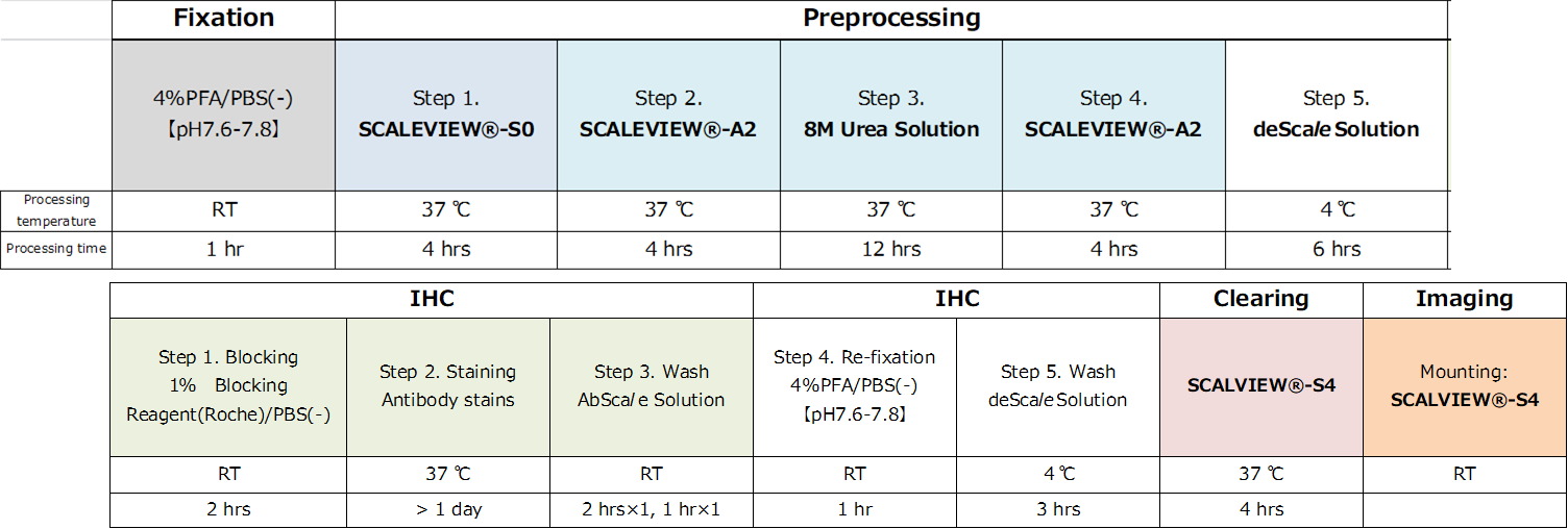
Protocol for clearing of brain slices (thickness: 1-2mm)
It is recommended to prepare slices after fixation process.
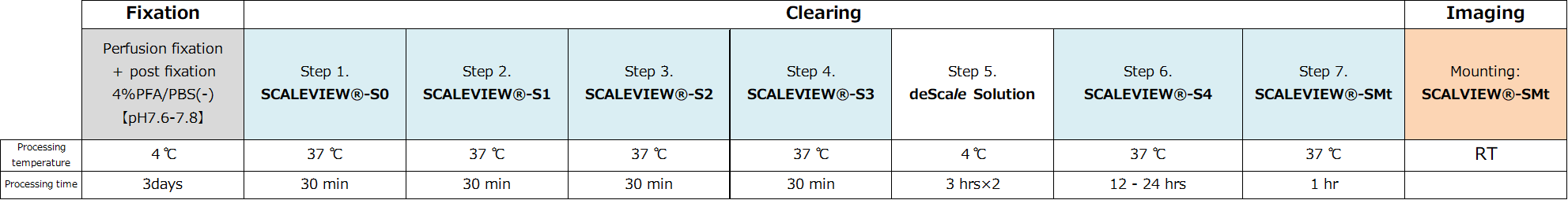
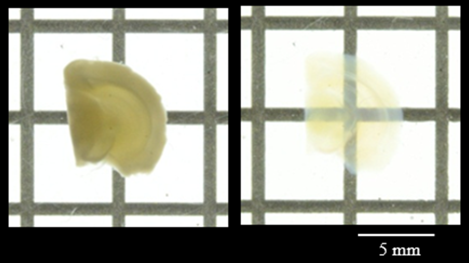 Figure 1. Transmittance images of mouse brain before and after clearing with SCALEVIEW-S Solutions.
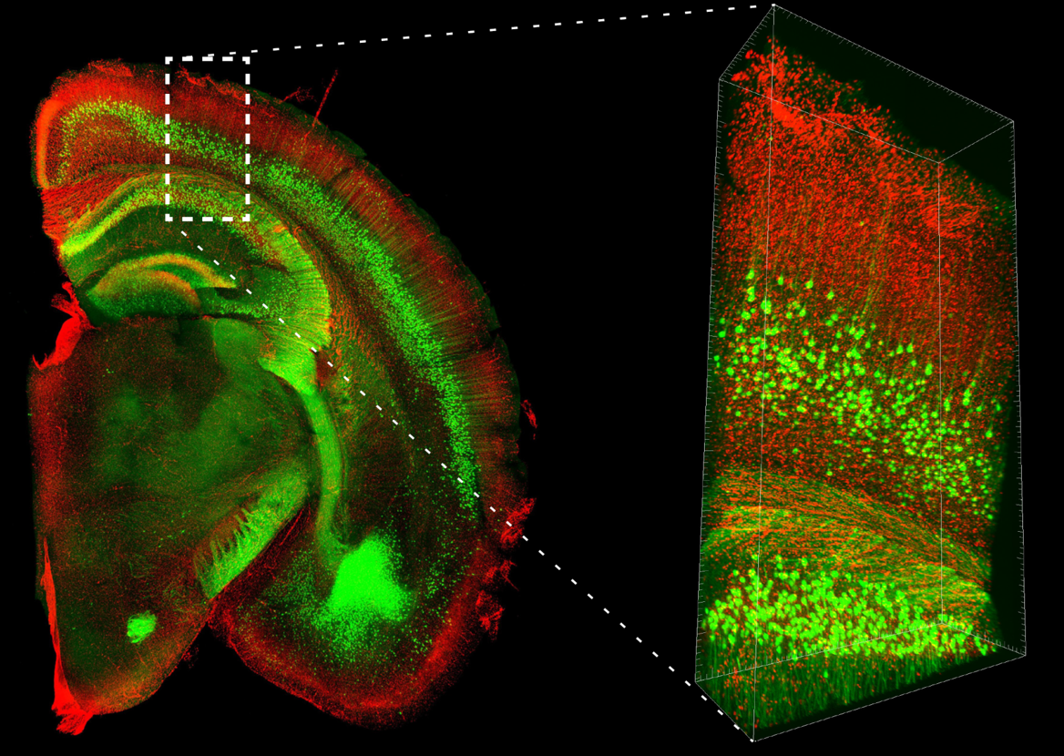
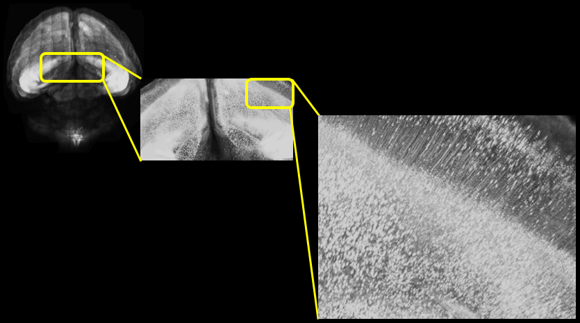 Figure 2. Two-photon microscope imaging of YFP-H line: Mouse whole brain
| Mouse | Thy1-YFP-H line, 20W, ♂ |
| Size | Whole |
| Microscope | Olympus FVMPE-RS |
| Objective lens | XLPLN10XSVMP (NA 0.6) |
| Laser | 960 nm (for YFP) |
| Image size | 512 x 512, 170 tiles, Z=8000 μm, Z Step16 μm |
Protocol for 3-D IHC (AbScale)
It is recommended to prepare slices after fixation.
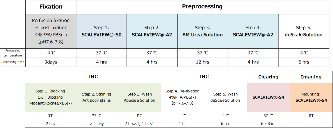
**AbScale Solution:0.33 M Urea and 0.1 % (wt/vol) Triton X-100 in PBS(-) Solution
Iba1 (RF635: Green) Amyloid-β (Alexa Fluor 488: Red) Tomato lectin (Texas Red: Blue)
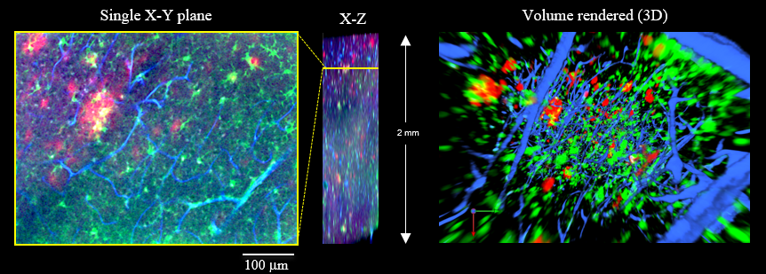 Figure 3. 3D visualization of Aβ plaques (red), microglias (green) and blood vessels (blue) from a 17-month-old AD model mouse.
| Microscope (CLSM) | Olympus FV1200 |
| Objective lens | XLPLN10XSVMP (NA 0.60) |
Protocol for 3-D Chemical Staining (ChemScale)
It is recommended to prepare slices after fixation.
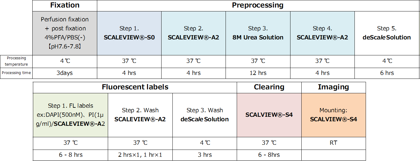
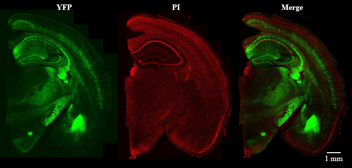 Figure 4. Confocal laser scanning microscope imaging of fluorescent labeled (PI) YFP-H line mouse brain slice (2 mm thick).
| Mouse | Thy1-YFP-H line, 42W, ♂ | Microscope (CLSM) | Olympus FV3000 (Inverted) |
| Size | Coronal Slice (2 mm) | Objective lens | UPLSAPO10x2 (NA 0.40) |
| Laser | 488 nm (for YFP), 561 nm (for PI ) |
Protocol for AbScale of neurosphere
 *AbScale Solution:0.33 M Urea and 0.1 % (wt/vol) Triton X-100 in PBS(-) Solution
** After clearing, embed and immobilize with 1.5% (wt/vol) agarose.
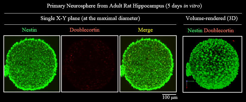
Figure5. 3D visualization of Neurosphere
| Microscope (CLSM) | Olympus FV1000 |
| Objective lens | UMPLFLN10XW (NA 0.3) |
References 参考文献
- Hama,H.et al. : Nature Neuroscience 14, 1481(2011).
- Hama,H.et al. : Nature Neuroscience 18, 1518(2015).
- Hama H, et al. : Protocol Exchange (2016), doi:10.1038/protex.2016.019
- Molly E, Boutin, et al :Scientific Reports, 8, 11135 (2018).